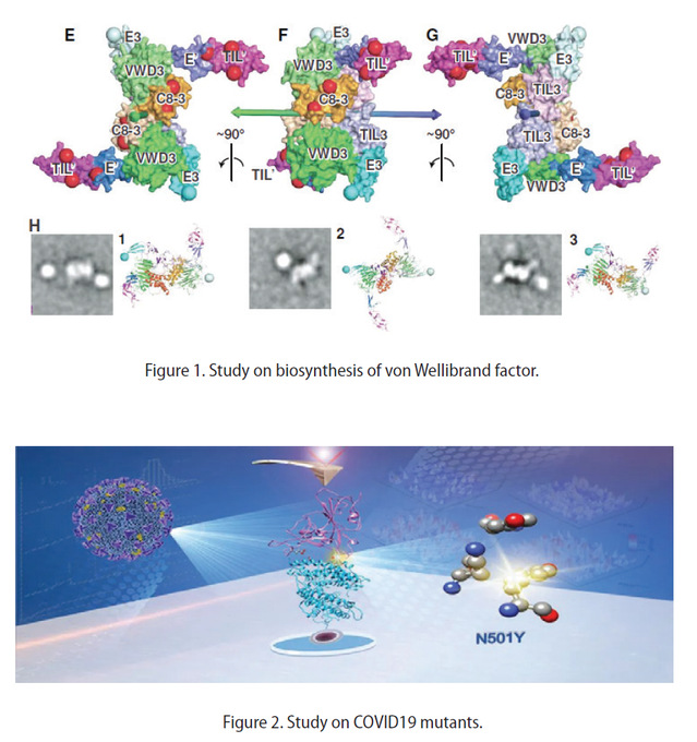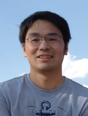
ResumeXianchi received his Ph.D. degree in Biochemistry and Molecular Biology (2004-2010) from the Shanghai Institutes for Biological Sciences, Chinese Academy of Sciences. His Ph.D. work was carried out in Dr. Jianping Ding’s lab focused on structural biology. From 2010 to 2018, Xianchi worked with Dr. Timothy Springer at Immune Disease Institute, Harvard Medical School as a postdoc and an instructor. In 2018, he started a Principal Investigator position in the School of Life Science of Nanjing University. Research FieldsProtein Biochemistry in Immune and Coagulation System Biosynthesis of von Willebrand factor Von Willebrand factor (VWF) monomers dimerize through their C-terminal domain in the endoplasmic reticulum (ER) and then through the N-terminal D'D3 assembly of mature VWF in the Golgi. The unusual process of disulfide bond formation in the Golgi enables formation of the ultralong, tail-to-tail, headto- head concatemers required for VWF activation in hemostasis. C-terminallytruncated VWF fragments are secreted as mixtures of monomers and dimers; the monomers contain two free cysteines, Cys-1099 and Cys-1142, which were proposed to form the dimerizing inter-chain disul de bonds. However, chemical determination of disul de bonds in VWF is challenging and only the Cys-1142/ Cys-1142' disul de has been con rmed. Surprisingly, a recent VWF D’D3 monomer crystal structure showed burial of Cys-1099 and Cys-1142, thus revealing how these residues are protected from disul de bond formation in the ER, but little about disulfide bond formation in the Golgi. Gel-forming mucins contain D assemblies homologous to those of VWF and also form multimers in the Golgi; however, in mucin MUC2, homologues of VWF Cys-1142 and Cys-1097 but not Cys-1099 formed dimerizing disul des. Multiple explanations for the discrepancy were proposed, but not resolved. In this study, we present evidence suggesting that disul de exchange between three Cys residues in VWF frees Cys-1097 to form a dimerizing disul de bond. Mutation of Spike Protein in SARS-CoV-2 Strengthens the binding to its Receptor SARS-CoV-2 is spreading around the world for the past year. Recently, several variants such as B.1.1.7, B.1.351, and P.1, sharing a key mutation N501Y on the RBD, appear to be more infectious to humans. To understand the underlying mechanism, we preformed cell surface binding assay, kinetics study, singlemolecule technique, and computational method to investigate the interaction between these RBD (mutations) and ACE2. Remarkably, RBD with the N501Y mutation exhibited a considerably stronger interaction, with a faster association rate and slower dissociation rate. Consistently, atomic force microscopy-based single-molecule force microscopy quantify their strength showing a higher binding probability and unbinding force for the mutation. Molecular dynamics simulations of RBD-ACE2 complexes indicated that the N501Y introduced additional - and -cation interaction for the higher force/interaction. Taken together, we suggested that the reinforced interaction from N501Y mutation in RBD should play an essential role in the higher transmission of COVID-19 variants.
Part-time Academic JobWork ExpericeAchievement1. Dong X. Springer T.A. (2021) Disulfide exchange in multimerization of von Willebrand factor and gel-forming mucins Blood 137(9), 1263-1267 2. Tian F$, Tong B$*, Sun L, Shi S, Zheng B, Wang Z, Dong X*, Zheng P*. “N501Y mutation of spike protein in SARS-CoV-2 strengthens its binding to receptor ACE2”, Elife, (2021) Awards |